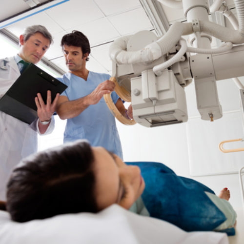
This year’s celebration will take place the week of November 3-9, marking the 124th year since the discovery of X-rays by Wilhelm Conrad Roentgen. The first National Radiologic Technology Week, (NRTW), was celebrated in July of 1979, and was organized by the American Society of Radiologic Technologists (ASRT). It was then changed to the week of November 8th to coincide with the day Roentgen made his discovery. It is now celebrated annually to highlight the important role medical imaging and radiation therapy have in the treatment of patients. Founded in 1920, the ASRT is the world’s largest and oldest radiologic science organization boasting a membership of over 94,000 people. The ASRT provides educational opportunities, promotes career opportunities, as well as establishing standards of practice, education and entry-level requirements.
This year’s theme is “waves of the future”. Before turning our attention to the future, I thought it would be interesting to take a look at the past and see where medical imaging had its origins and see how far we have advanced since 1895. The timeline provided below was copied from the website www.sciencelearn.org.nz. I have highlighted what I consider to be some of the more important events in the history of medical imaging.
November 8, 1895 – X-rays discovered
X-rays are discovered by German physicist Wilhelm Roentgen. He took the first X-ray image, showing the skeletal composition of his wife’s left hand.
1946 – Nuclear Magnetic Resonance (the predecessor to MRI)
American physicists Edward Purcell and Felix Bloch independently discover nuclear magnetic resonance (NMR).
1955 – Ultrasound for medical diagnosis
Ian Donald, a Scottish physician, begins to investigate the use of ultrasound to diagnose gynaecological patients.
1967 – CT scanning conceived
Godfrey Hounsfield, an English electrical engineer, conceives the idea for computed tomography.
1971 – First CT scan of patient’s brain
The first CT scanner is installed in Atkinson Morley Hospital, Wimbledon, England, and the first patient brain scan is performed on October 1, 1971.
1974 – PET camera developed
Michael Phelps develops the first positron emission tomography (PET) camera and the first whole-body system for human and animal studies.
July 3, 1977 – First human MRI body scan
The first MRI body scan is performed on a human using an MRI machine developed by American doctors Raymond Damadian, Larry Minkoff, and Michael Goldsmith.
Early 1980s – MRI in hospitals
MRI scanners are installed in hospitals.
1990s – Prenatal ultrasound becomes routine
Ultrasound has become a routine procedure in pregnancy to check the baby’s development and to pick up any abnormalities.
2000 – Medical invention of the year
Time Magazine names the PET/CT scanner the medical invention of the year.
In Conclusion
There really have been some amazing developments over the past 124 years. The invention of x-rays not only allows us to diagnose diseases, but it also allows us to treat them. The field of radiation therapy followed immediately after the discovery of x-rays. This is a field that has had much growth over the decades; moving from high dose long exposure to the now commonplace fractional dosing, maximizing tumor destruction and salvaging normal tissue. I think that Wilhelm Roentgen would be very pleased with how his invention has bettered society.
To all of you that work as a radiologic technologist or in a support service role, I wish you a Happy National Radiologic Technologist week!
If you are a radiologic technologist or radiation therapy technologist that is looking for employment or you are in need of a technologist to fill an opening please visit our website, www.bartonhealthcarestaffing.com to see how we can help.

